Artificial intelligence in the laboratory PMA: emerging applications and implications for process quality
Artificial intelligence (AI) is gradually finding its way into the practice of medically assisted reproduction (MAP), changing the methods of acquisition, analysis and integration of data biological data. The main focus lies in the possibility of transform traditionally qualitative and operator-dependent assessments into quantitative, standardisable information and reproducible (Salih et et al., 2023). The The first applications were focused focused on computer vision and the morphological assessment of embryos; subsequently, AI has been extended to the analysis of embryonic development dynamics, the assessment of gametes and support for various stages of the laboratory process.
Assessment of morphology and of development embryonic
Embryonic selection is the area of application of theAI in human embryology. Convolutional neural networks (CNNs) enable the analysis of embryonic images by identifying complex morphological patterns. The systematic review by Salih et al. highlighted favourable performance of AI systems, but also a high degree of methodological heterogeneity, with prevalence of studies retrospective and datasets often single-centre (Salih et al., 2023).
A useful application for laboratories without time-lapse systems is DeepEmbryo. Borna and al. have developed a model based on CNN and transfer learning using static images of the embryo, acquired at approximately 19, 43 and 67 hours
post-insemination (Borna et al., 2024). The model has achieved an’accuracy of up to of 75 per cent in predicting pregnancy outcome, which is higher than that achieved using a single image. The use of sequential images therefore allows information on the dynamics of embryonic development to be incorporated, without requiring continuous monitoring.
A further area concerns the probabilistic prediction of euploidy. ERICA (Embryo Ranking Intelligent Classification Algorithm), developed by Chavez-Badiola et al., uses static images of blastocysts to identify characteristics associated with the probability of euploidy and implantation (Chavez-Badiola et al., 2020). In the dataset of 1,231 images, the model achieved 70 per cent accuracy in predicting euploidy status. Of the embryos classified as euploid, 79 per cent were found to be euploid upon subsequent genetic analysis. Furthermore, a euploid embryo was selected as
first choice in 78.9 per cent of cases, and at least one euploid embryo was included amongst the top two in 94.7 per cent of cases. However, these results must be interpreted with caution: the analysis of images does not determine directly the chromosomal but estimates the probability of euploidy based on morphological characteristics. AI prediction of ploidy is therefore not equivalent to a genetic diagnosis, but may serve as a tool to support embryo selection.
A recent recent development marked by the introduction of models multimodal. Wang and al. have developed IVFormer, capable of integrating embryonic images, time-lapse sequences obtained via time-lapse and clinical information (Wang et al., 2024). This approach views embryonic development as a dynamic process, integrating morphology, developmental kinetics and clinical characteristics into the assessment of the outcome.
From the embryo to gametes: new applications in the laboratory
The use ofAI is not limited to embryo selection, but also involves other activities in the laboratory for assisted reproduction. Goss et al. have developed a system for identify spermatozoa in surgical specimens from patients with non-obstructive azoospermia (Goss et al., 2024). In the simulation of clinical use, the AI has reduced the average time spent for searching for sperm from approximately 169 to 99 seconds. Thevalue of this application therefore lies both in the identification of rare spermatozoa and in the reduction of a labour-intensive stage and
operator-dependent, whilst keeping the embryologist responsible for verification final verification.
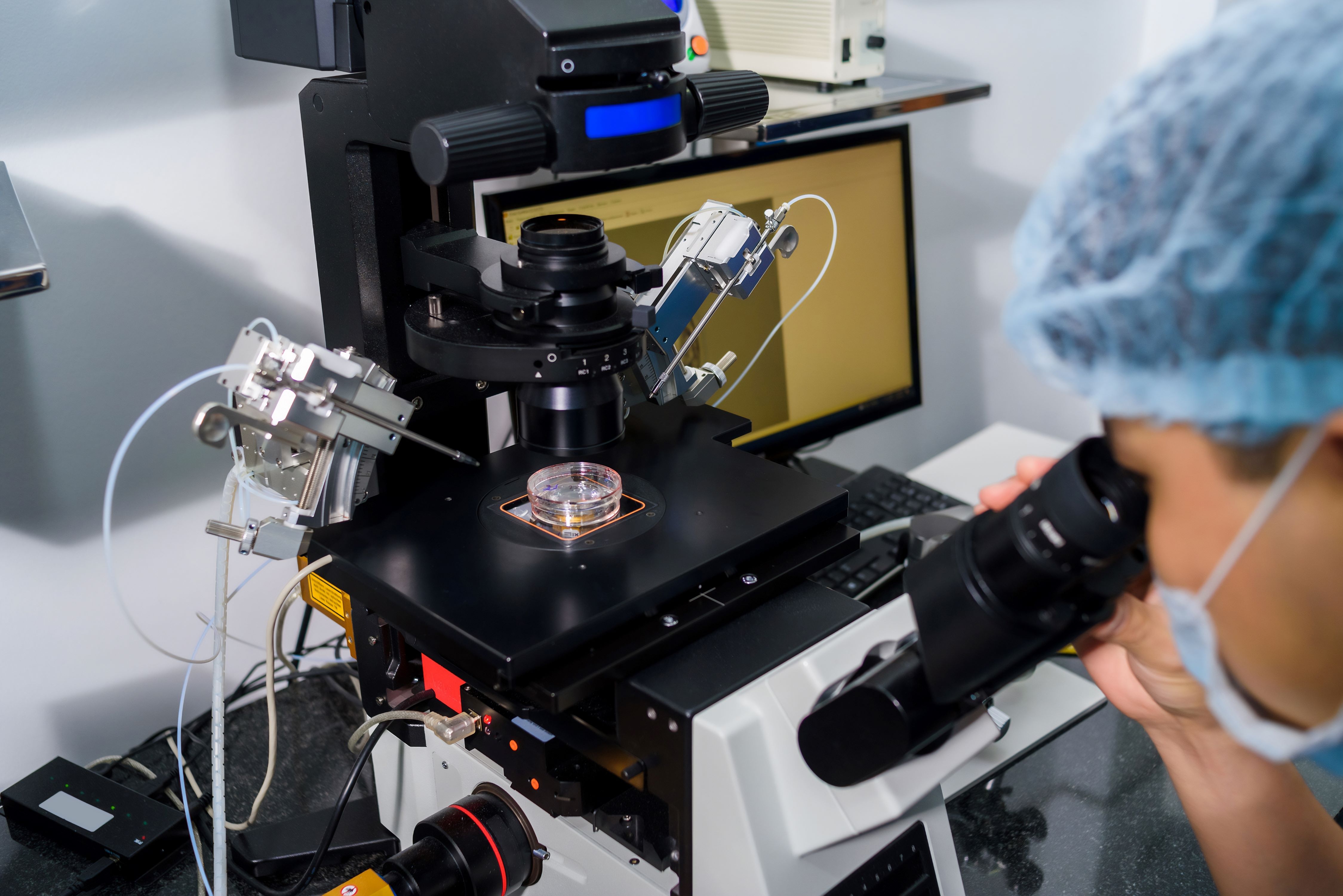
Other possible applications include gamete classification and assistance with micromanipulation and ICSI, the analysis automated image processing, theprocessing of data produced by time-lapse systems and support for the management of laboratory workflows (ASRM & SRBT, 2026).
From algorithmic performance to clinical validation
An aspect fundamental in the interpretation of literature is the distinction between predictive performance and clinical benefit. An algorithm may, in fact, demonstrate high classification capabilities without necessarily leading to an improvement in outcome.
One particularly relevant is the multicentre randomised by Illingworth et al. (2024), which involved 1,066 patients across 14 centres and compared embryo selection using the iDAScore deep learning system with selection based on morphology conventional The rate of clinical pregnancy is the result to 46.5 per cent in the group
AI and to 48.2 per cent in the conventional group; the pre-established criterion of not inferiority was not therefore achieved (Illingworth et al., 2024).
These results highlight how the introduction of AI cannot be based solely on algorithmic accuracy, but requires validation across populations and contexts that are and, above all, the demonstration of a genuine clinical benefit.
Conclusions
AI is evolving from a tool for automated image analysis into a technology with the potential to be applied to various stages of PMA laboratory process, from selection embryo selection to gamete assessment and the management of laboratory processes.
Its value lies does not lie in replacing the embryologist, but in the possibility to to complement this with tools capable of reducing variability and increasing standardisation during critical stages. AI can also contribute to organisational efficiency by reducing and promoting a more quantitative monitoring of performance. Its introduction, however, requires a cautious approach, based on prospective validation, verification of reproducibility and of safety and demonstration of a genuine clinical utility beyond mere predictive accuracy.
The future of the ART laboratory could therefore be represented by a model
AI-assisted, in which technology, expertise of the embryologist and quality assurance system are integrated to make processes more standardised, traceable, efficient and reproducible.
Bibliography
1. American Society for Reproductive Medicine; Society of Reproductive Biologists and Technologists. Artificial intelligence in the in vitro fertilisation laboratory: a committee opinion. Fertil Steril. 2026. doi:10.1016/j.fertnstert.2026.05.004.
2. Borna MR, Sepehri MM, Maleki B. An artificial intelligence algorithm to select the most viable embryos, taking into account current processes in IVF laboratories. Front Artif Intell. 2024;7:1375474. doi:10.3389/frai.2024.1375474.
3. Chavez-Badiola A, Flores-Saiffe-Farías A, Mendizabal-Ruiz G, Drakeley AJ, Cohen J. Embryo Ranking Intelligent Classification Algorithm (ERICA): an artificial intelligence clinical assistant for predicting embryo ploidy and implantation. Reprod Biomed Online. 2020;41(4):585–593. doi:10.1016/j.rbmo.2020.07.003.
4. Goss DM, Vasilescu SA, Vasilescu PA, Cooke S, Kim SH, Sacks GP, et al. Evaluation of an artificial intelligence-facilitated sperm detection tool in azoospermic samples for use in ICSI. Reprod Biomed Online. 2024;49(1):103910. doi:10.1016/j.rbmo.2024.103910.
5. Illingworth PJ, Venetis C, Gardner DK, Nelson SM, Berntsen J, Larman MG, et al. Deep learning versus manual morphology-based embryo selection in IVF: a randomised, double-blind non-inferiority trial. Nat Med. 2024;30(11):3114–3120. doi:10.1038/s41591-024-03166-5.
6. Salih M, Austin C, Warty RR, Tiktin C, Rolnik DL, Momeni M, et al. Embryo selection using artificial intelligence versus embryologists: a systematic review. Hum Reprod Open. 2023;2023(3):hoad031. doi:10.1093/hropen/hoad031.
7. Wang G, Wang K, Gao Y, Chen L, Gao T, Ma Y, et al. A generalised AI system for human embryo selection covering the entire IVF cycle via multi-modal contrastive learning. Patterns (NY). 2024;5(7):100985. doi:10.1016/j.patter.2024.100985.